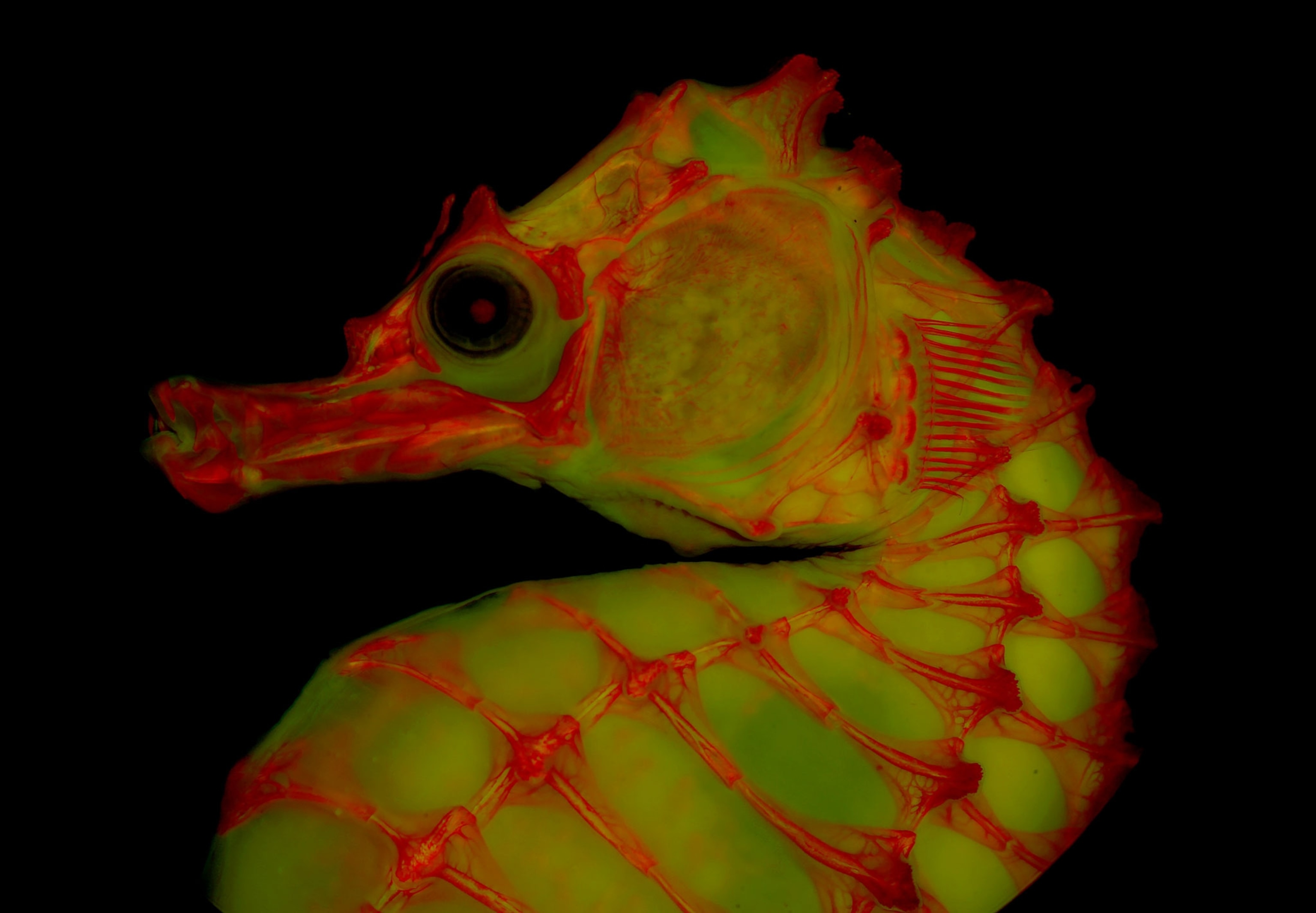
Fluorescent light, red dye, and gelatin are the ingredients of an emerging photography technique that allows scientists to better visualize the skeletons of animals.
Researchers who study vertebrates have long relied on what’s known as clearing and staining—stripping specimens of their soft tissues and coloring the remains with red dye—to take detailed images, used to examine anatomy and the relationships among species. But stripped of ligaments and musculature, skeletons can be flaccid, making them difficult to prop up and photograph from certain angles.


“There's so many different pictures you just can't get,” says Leo Smith, an ecology and evolutionary biology professor at the University of Kansas, who helped develop the new technique. “If it’s a catfish, it’s going to sit on its stomach, and that’s all you got. If it’s a trout or something, it’s going to lie on its side because it will just collapse in another dimension.”
That’s where the gelatin comes in. Its jelly-like texture can hold skeletons in a pose, allowing them to be photographed at different angles, and then washes clean off when the photo shoot is done. Combined with red dye and illuminated with fluorescent light, the method yields opportunities to take images that were once impossible.
Glowing red
On a late night in 2013, Smith, the lead author of the 2018 paper describing the technique, placed a dye-stained fish skeleton under a fluorescence microscope—which uses a high-intensity light instead of visible white light—on a whim.
“I just stuck it under there, and I was like, Holy cow, this is amazing,” says Smith, ”because the fluorescence really brought out detail.”

“It’s very similar to glow-in-the-dark toys,” says Matt Davis, a biology professor at St. Cloud State University, in Minnesota, and a co-author of the paper. “The principle is basically the same. It’s absorbing light and then re-emitting it.” (Platypuses, flying squirrels, sea turtles, and other creatures are naturally biofluorescent.)
The beauty of fluorescence imaging, Smith says, is that it declutters aspects of the specimen, allowing researchers to pay attention to details they didn’t or couldn’t notice before.
The gelatin portion of the method was refined by Chesney Buck, a volunteer who assisted in Smith’s lab, and Matt Girard, a doctoral student who works under Smith at the University of Kansas. Girard says embedding dyed specimens in gelatin opened a new realm of possibilities.
“When you can actually move something or put a pair of tweezers in there, or hold it with your hand and move it, you can start to see how bones articulate with each other,” he says. “Or you can see if there's something behind a bone, because a lot of things—maybe not in humans but in other animals—will have layers of bones.”


Serendipity
Smith, Davis, and Girard have begun to experiment with different wavelengths of light, camera filters, and microscopes to see what else the animals will reveal to them.
“We reconstruct these trees of life, and explore how [the specimens] evolve through time, and how they're all related to one another,” Davis says. “We do that by looking for shared features, which might be genetic, or they might be anatomical.”
But the rest of the job, Davis says, is about enjoying it. “Part of science is discovery, and part of it is also just having fun.” (See more amazing photos of cleared and stained fish here.)
After all, that’s how Smith discovered the technique in the first place. Any moment of serendipity, he says, could lead to another breakthrough.
Editor's note: This story was updated March 23, 2021, to correctly identify the stippled clingfish.


