These images reveal a world that can’t be seen with the naked eye

From the hairs on a sea buckthorn plant to a rodent’s optic nerve, the winners of Nikon’s Small World photo contest offer a fascinating glimpse into the microscopic.
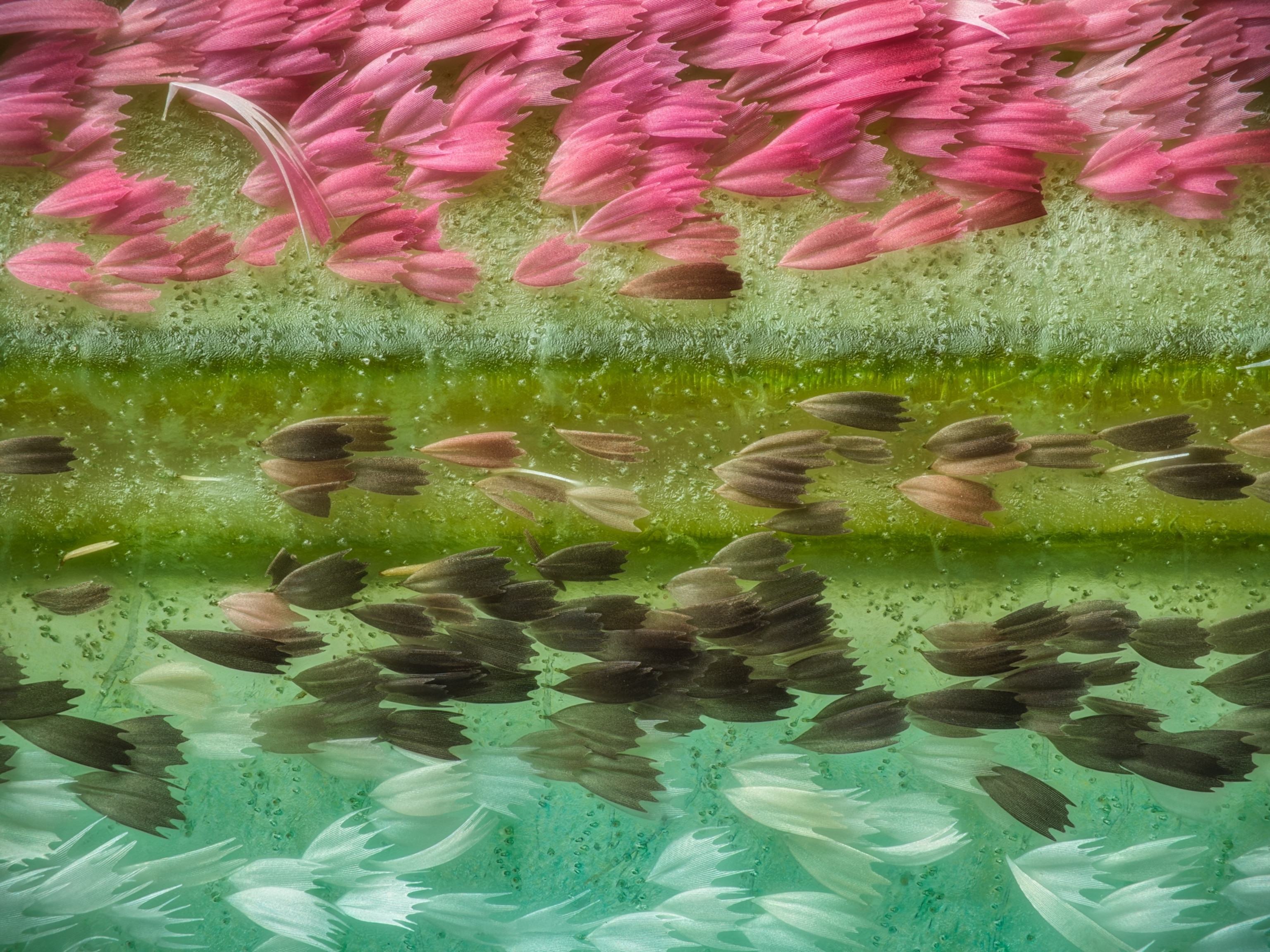
A microscopic view of scales on the wing of a moon moth; the scales are arranged in orderly rows, going from shades of pink, to shades of brown and green, to shades of white and blue.
Up close, the edge of a Chinese moon moth’s (Actias ningpoana) wing looks like cars driving on a road, says photographer Yuan Ji. 
Photograph by Yuan Ji, Nikon Small World
BySarah Gibbens
October 17, 2023
7 min read

Microscopic photography has the power to reveal the world hidden under a microscope. For 49 years, Nikon’s Small World Photo Microscopy Competition has been showcasing the best photos of our tiny world. 

This year’s winning photo was taken by researcher Hassanain Qambari with assistance from Jayden Dickinson from the Lion’s Eye Institute, a vision research center in Perth, Australia. It shows a rodent’s retina centered on the optic nerve, which transmits information between the eye and the brain. 

“[The photo] gives you a sense of what structures are at play and how much energy is used from the moment we open our eyes,” says Qambari. 

Streaks of red, yellow, and green reveal the molecular inner workings of this critical nerve, an unprecedented look that could help researchers understand how to treat a condition called diabetic retinopathy. In humans, this condition causes vision to blur and then disappear entirely. Early detection of the disease could help stop it in its tracks—and the better doctors can see inside the eye, the better they can understand its complex inner workings, insights that could save someone from blindness. 

It’s just one example of the kinds of fascinating new insights that can be discovered by looking at our world up close. Below is a selection of our favorite photos, selected for being exemplifying both art and science. 
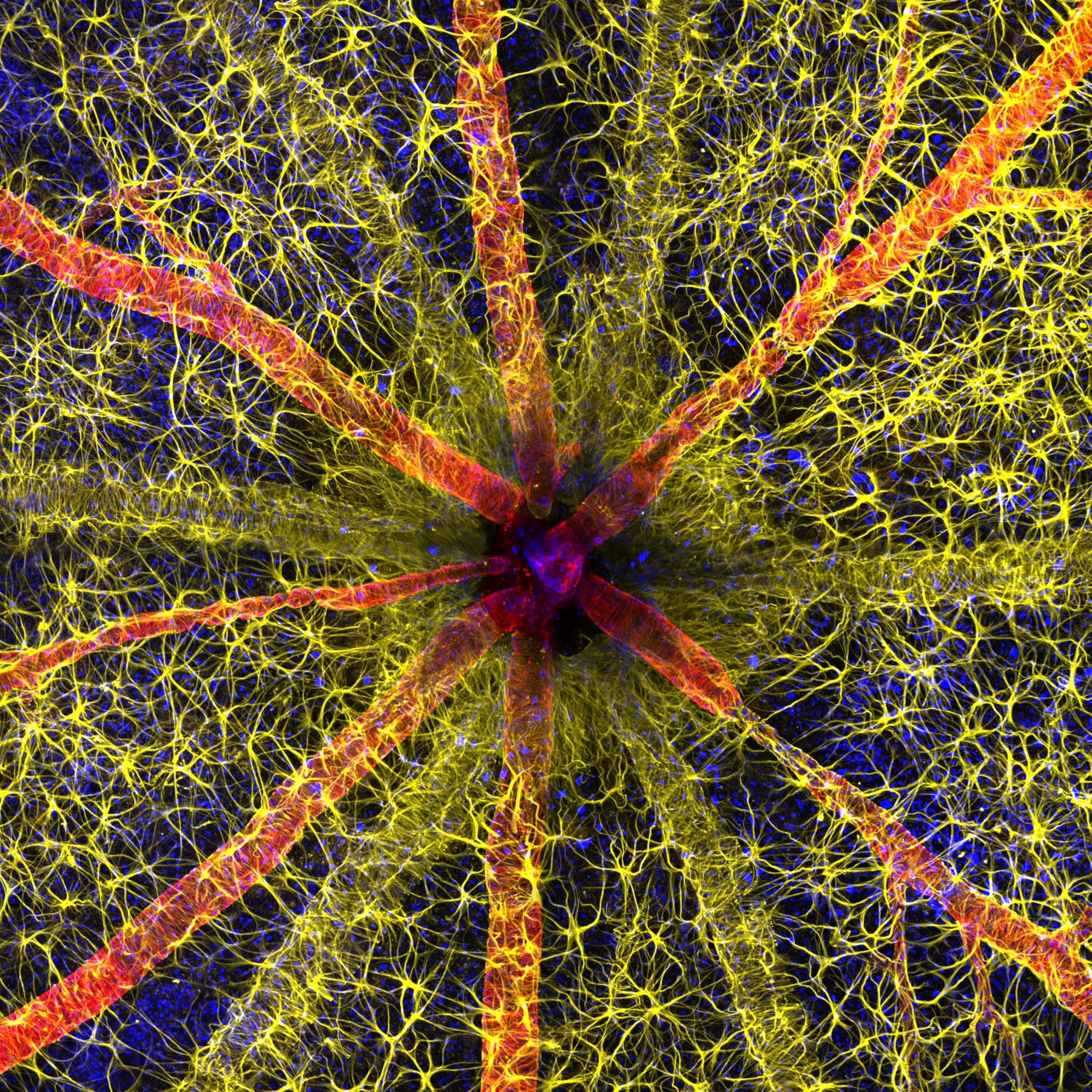
A rodent optic nerve head, showing red contractile proteins covered in yellow astrocytes, converging to a central point.
This year's winning image shows the head of a rodent's optic nerve. Since 2021, Qambari has been investigating methods for the early detection and reversal of diabetic retinopathy, a complication of diabetes that can cause permanent damage to the blood vessels of the retina.
Photograph by Hassanain Qambari & Jayden Dickson, Nikon Small World
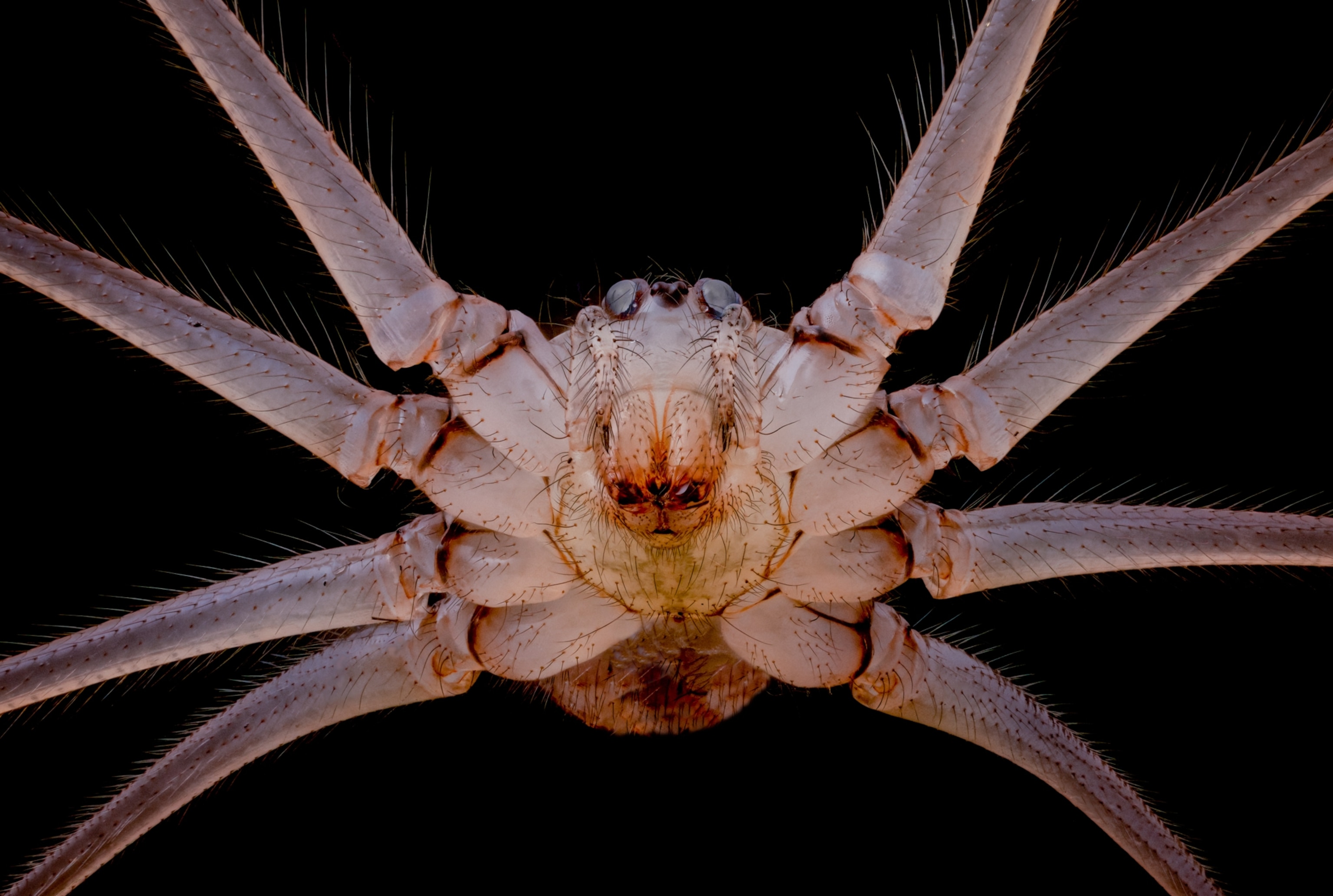
The underside of a cellar spider, which at 10X magnification almost appears as an alien's head.
The underside of a cellar spider (Pholcus phalangioides), also commonly called a daddy long legs. Last year, Andrew Posselt’s photograph of this same species won fourth place.
Photograph by Dr. Andrew M Posselt, Nikon Small World
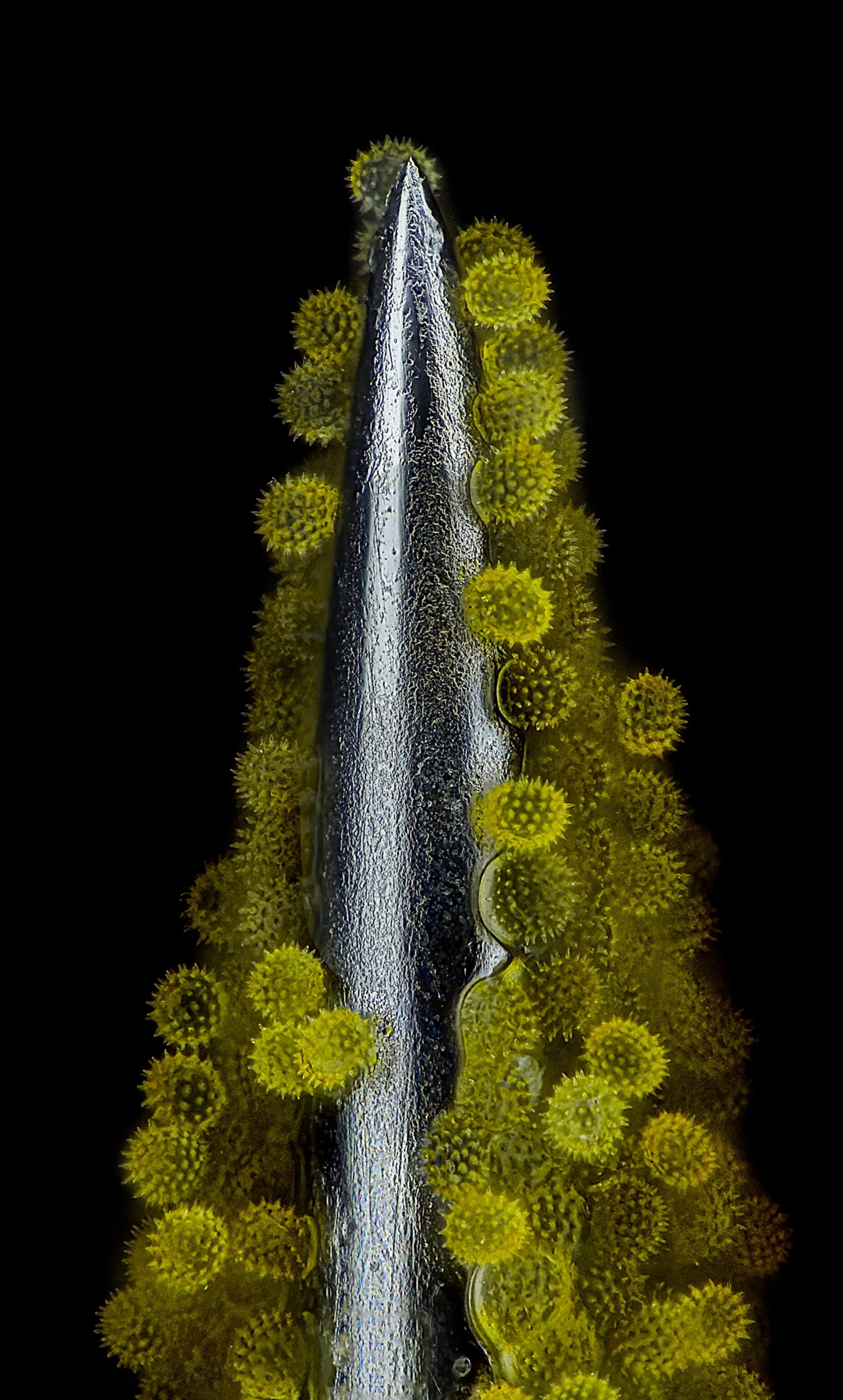
Spiky, yellow balls of pollen sticking to the metal point of an acupuncture needle
Lining the edges of an acupuncture needle are clusters of sunflower pollen. The spiky exterior helps pollen cling to plants. This image won 14th place.
Photograph by John-Oliver Dum, Nikon Small World
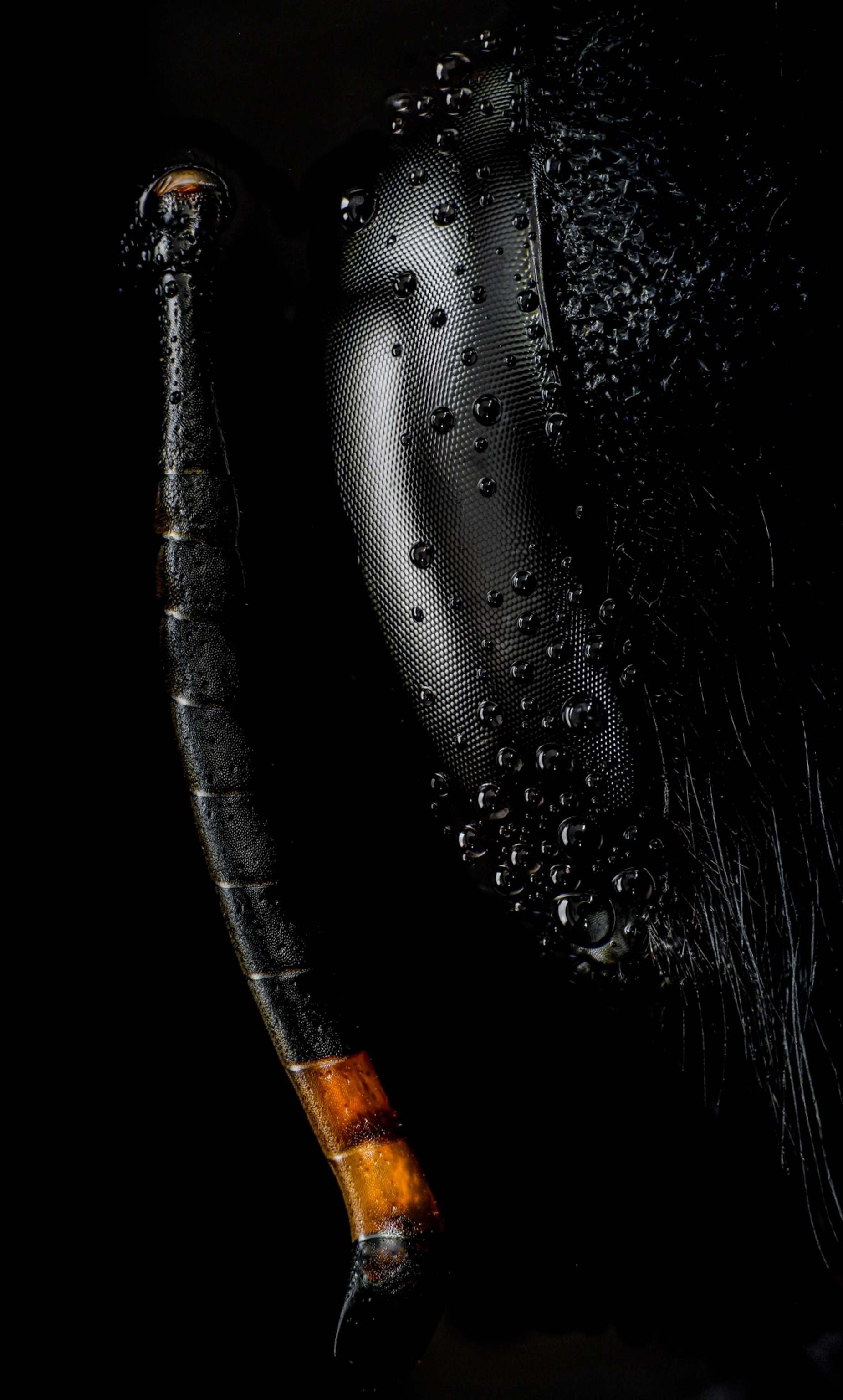
The head and antenna of a carpenter bee up close; droplets of liquid cling to the black textured eye of the bee.
The head and antenna of a carpenter bee (Xylocopa violacea).
Photograph by Ángel Navarro Gómez, Nikon Small World
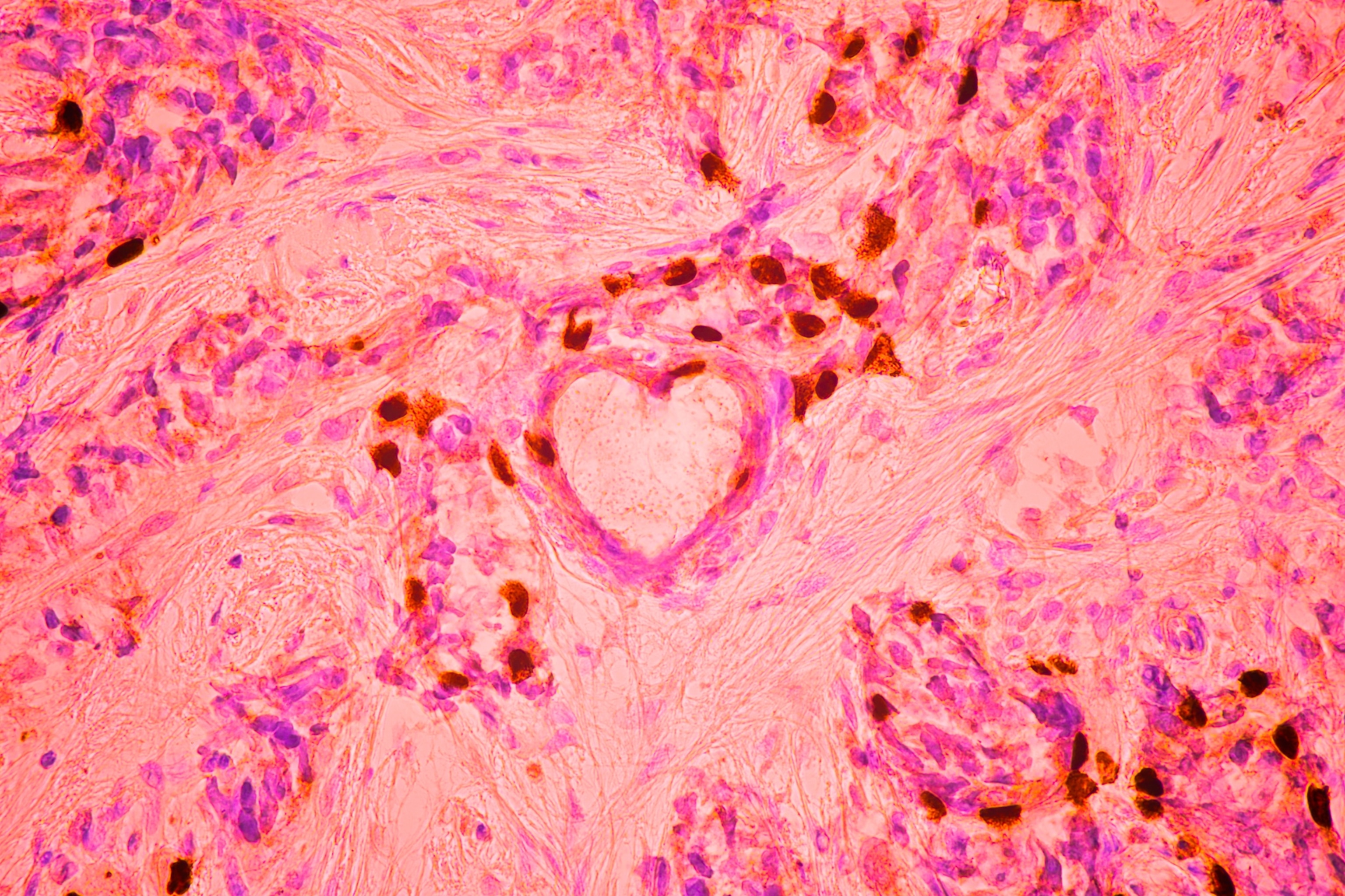
A microscopic view of breast cancer cells, which have coincidentally formed the shame of a heart
This microscopic view of breast cancer cells, which won third place, formed the shape of a heart.
Photograph by Malgorzata Lisowska, Nikon Small World
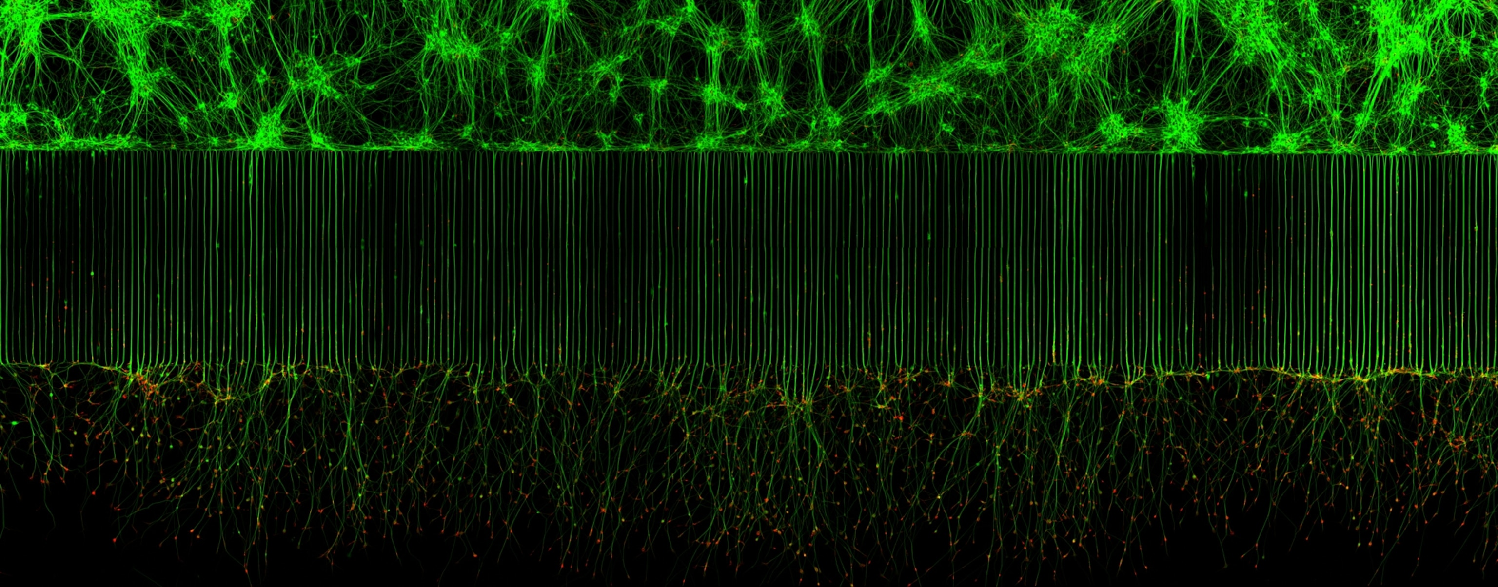
Motor neurons labeled to fluoresce green along the cell bodies, which starts as a complex web at the top of the frame, but organizes into neat vertical lines by the middle and bottom of the image.
An up-close look at motor neurons responsible for muscle movement. By observing these neurons at a cellular and molecular level, researchers can learn new therapeutic strategies to tackle neurodegenerative diseases such as ALS, or Lou Gehrig's Disease.
Photograph by Melinda Beccari & Dr. Don W. Cleveland, Nikon Small World
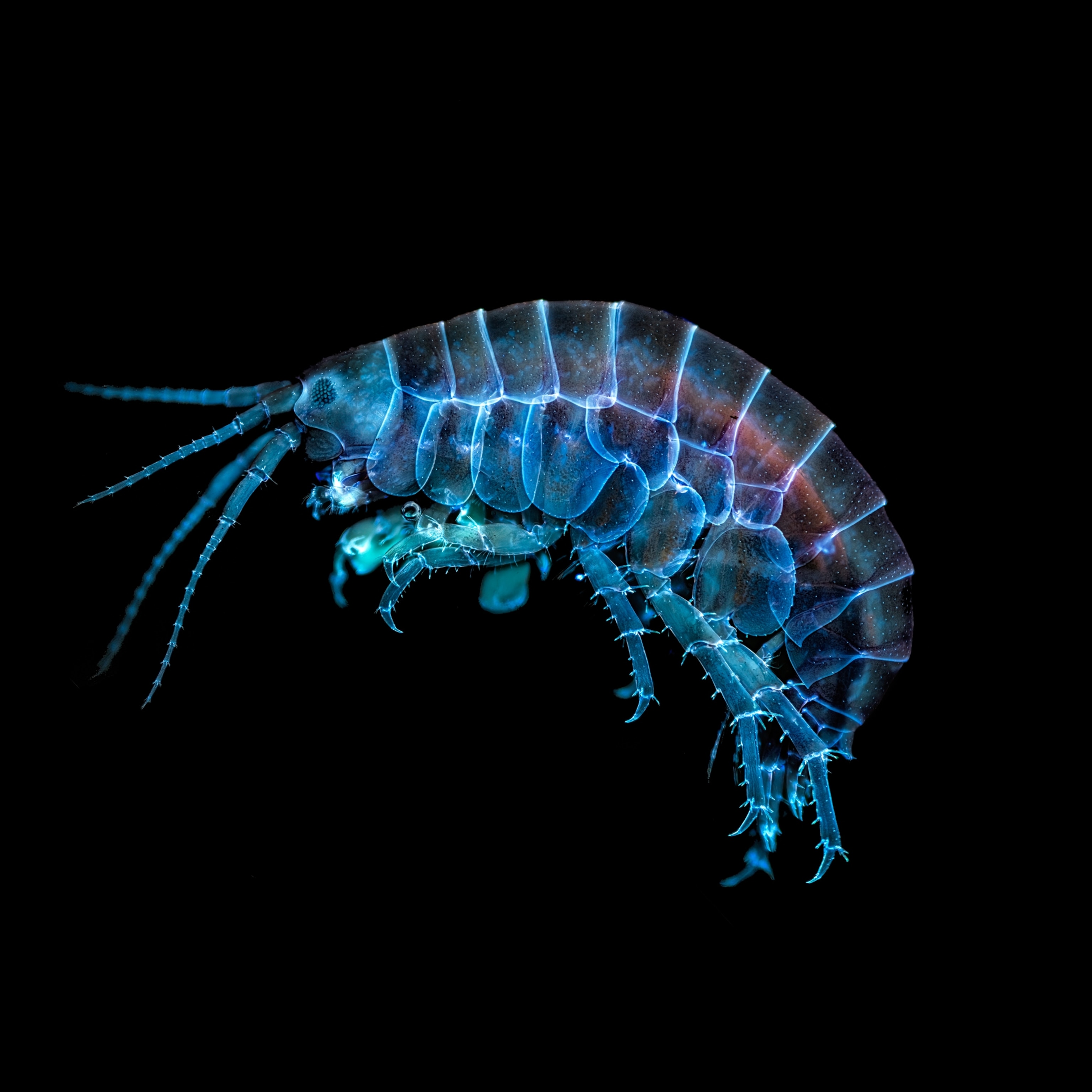
A microscopic view of an amphipod, which glows electric blue under UV light
Under a UV light, this freshwater amphipod is naturally fluorescent.
Photograph by Taylor Bell, Nikon Small World
A microscopic view of crystallized sugar syrup, which appears as intricately folded origami in shades of blue and white.
Under polarized light, crystallized sugar looks like the jagged, folded pages of a book.
Photograph by Dr. Diego García, Nikon Small World
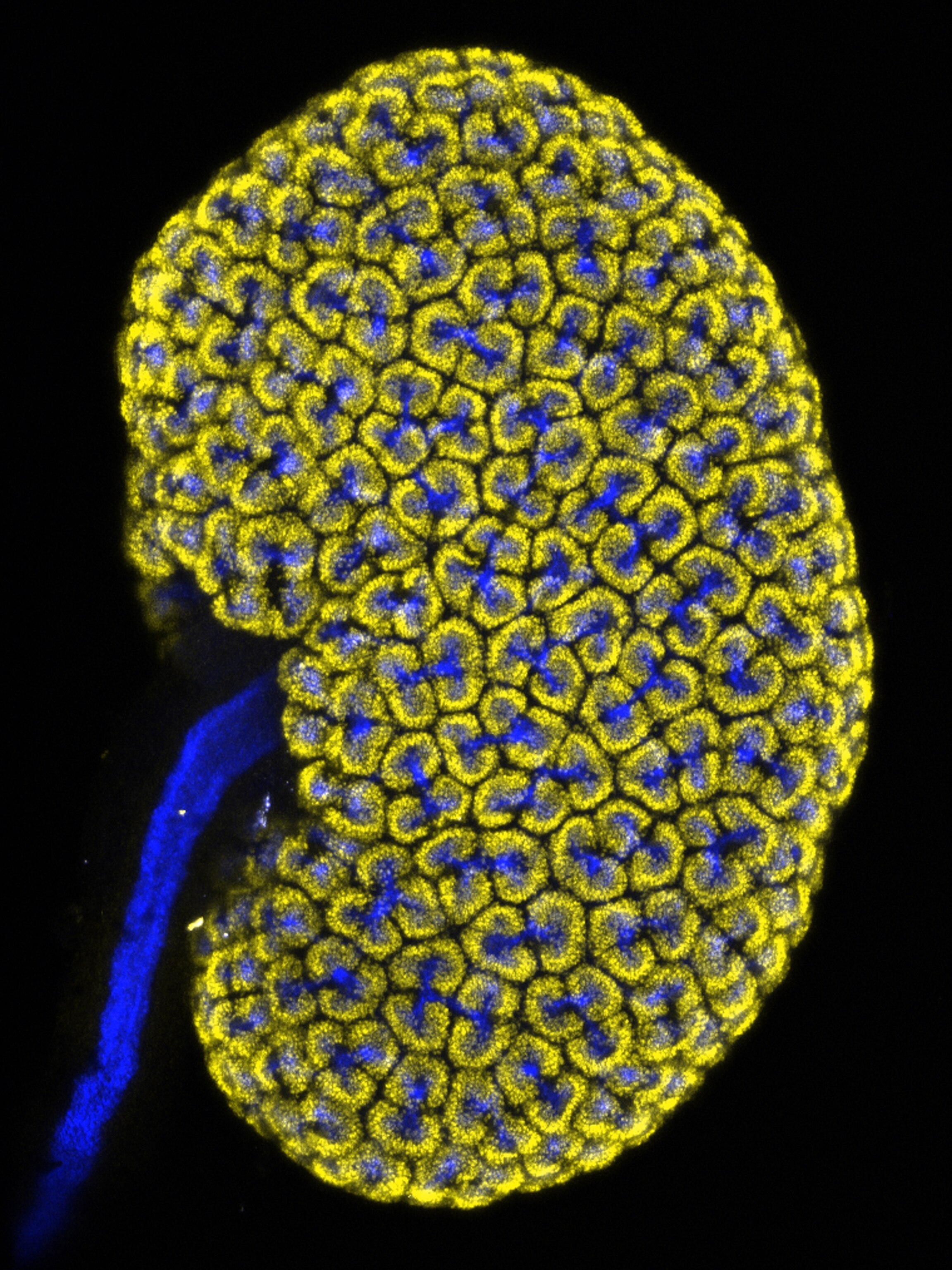
Embryonic mouse (Mus musculus) kidney showing the collecting duct (blue) and nephron progenitor (yellow) cells
The kidney of a mouse embryo shows clusters of two different and essential components that help this organ function properly.
Photograph by Dr. Lori O'Brien, Nikon Small World
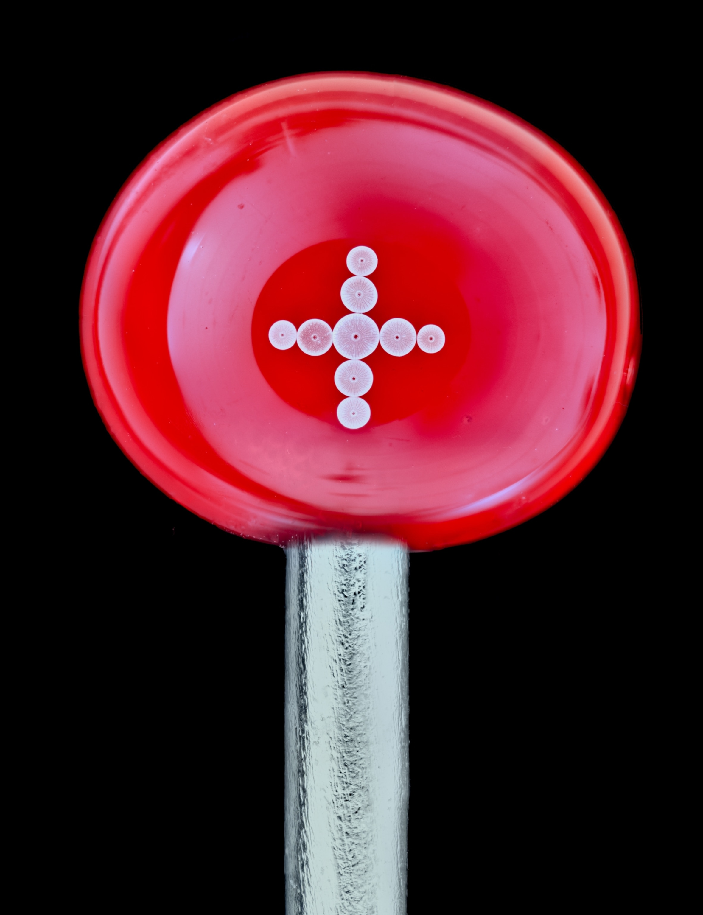
Diatoms (single-celled algae) arranged on the head of a pin
Diatoms are a type of single-cell algae found in every type of aquatic environment. To show just how tiny a diatom is, Jan Rosenboom placed one above the head of a pen. Arranging the diatom in the exact right position was a challenge—ultimately he moved it into place using a single strand of his own hair.
Photograph by Jan Rosenboom, Nikon Small World
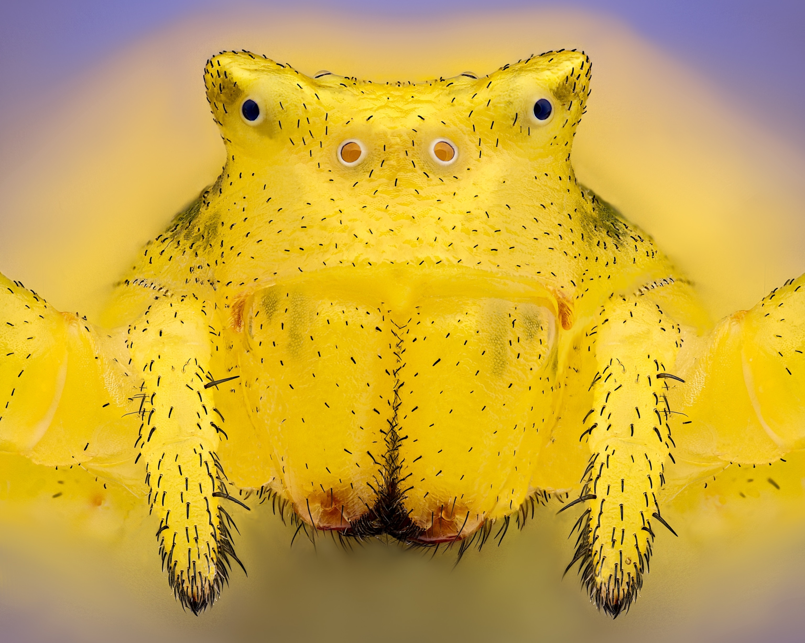
Crab spider (Thomisus onustus)
A close-up of a crab spider (Thomisus onustus). Female crab spiders can change their appearance from white, yellow, or pink to camouflage themselves in flowers.
Photograph by Sébastien Malo, Nikon Small World
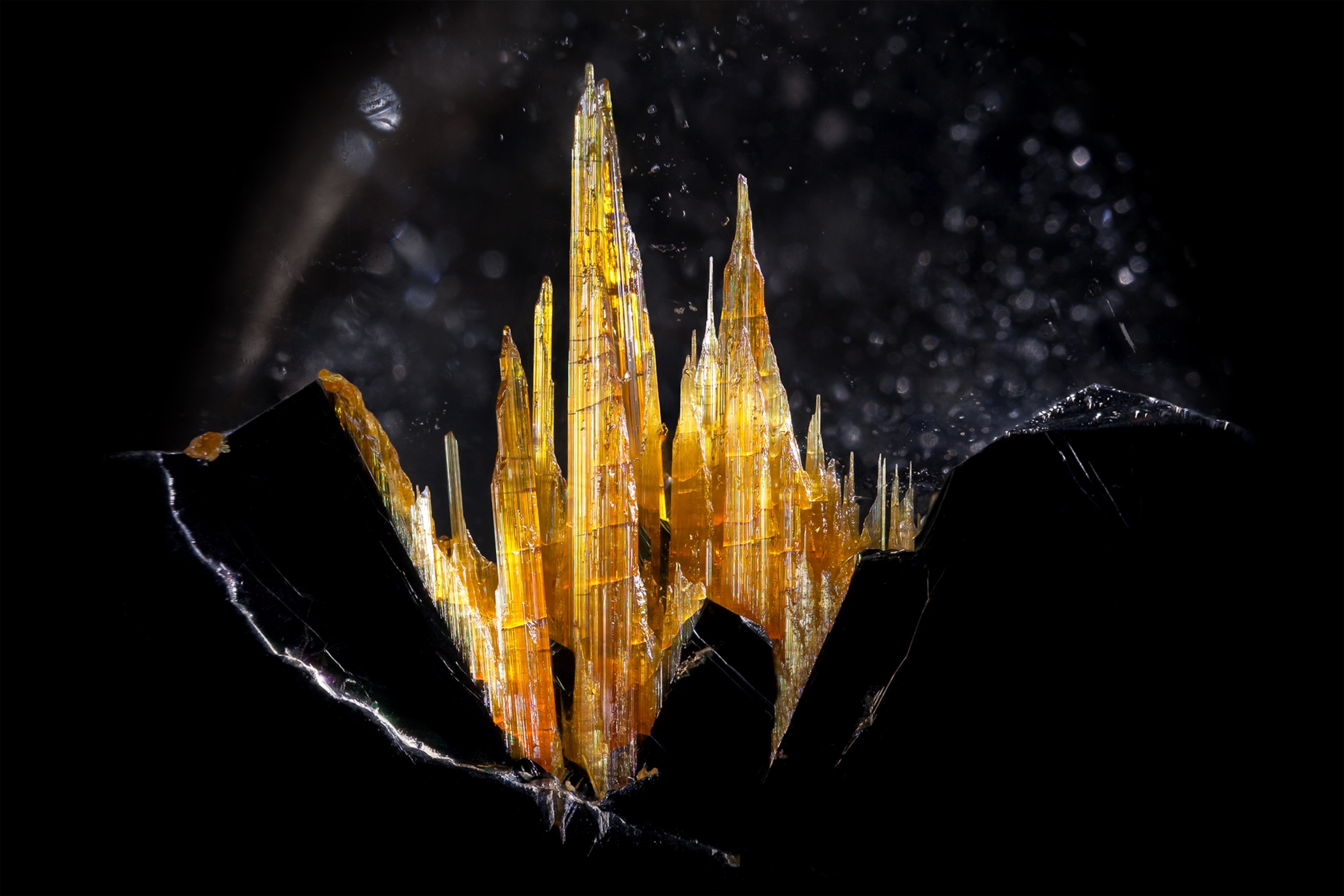
Golden rutile in quartz
Rutile is a type of mineral found in many rocks and often mined to produce titanium. Here, inside quartz, it appears golden.
Photograph by Danny Sanchez, Nikon Small World
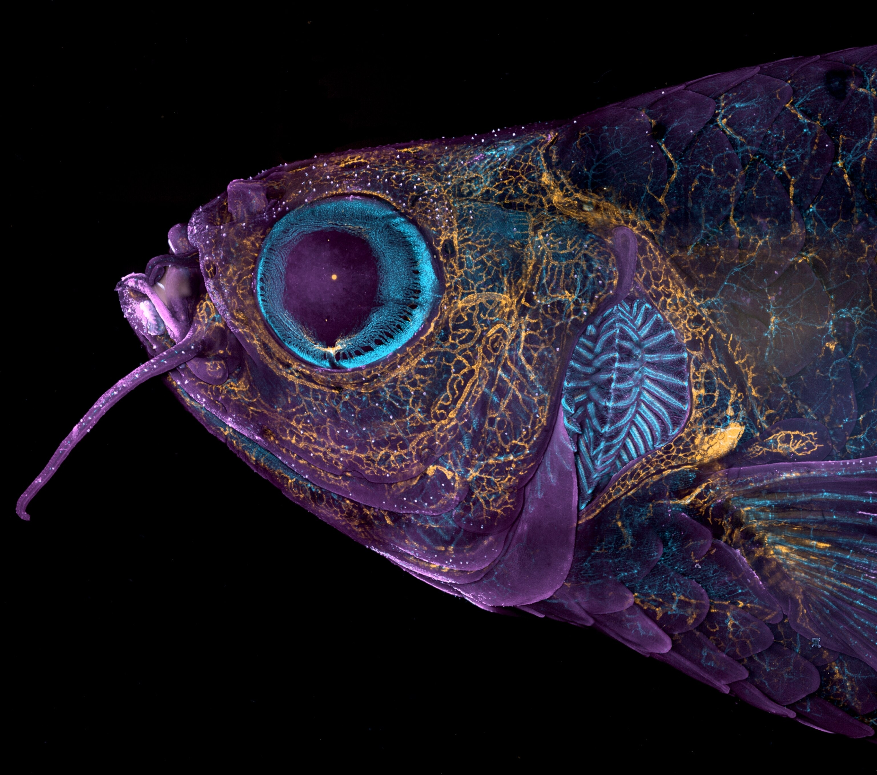
Adult transgenic zebrafish head showing blood vessels (blue), lymphatic vessels (yellow), and the skin and scales (magenta)
The head of an adult genetically modified zebrafish showing blood vessels (blue), lymphatic vessels (yellow), and the skin and scales (magenta). Images like these help researchers learn how animals like zebrafish grow and develop. 
Photograph by Daniel Castranova & Dr. Brant Weinstein, Nikon Small World
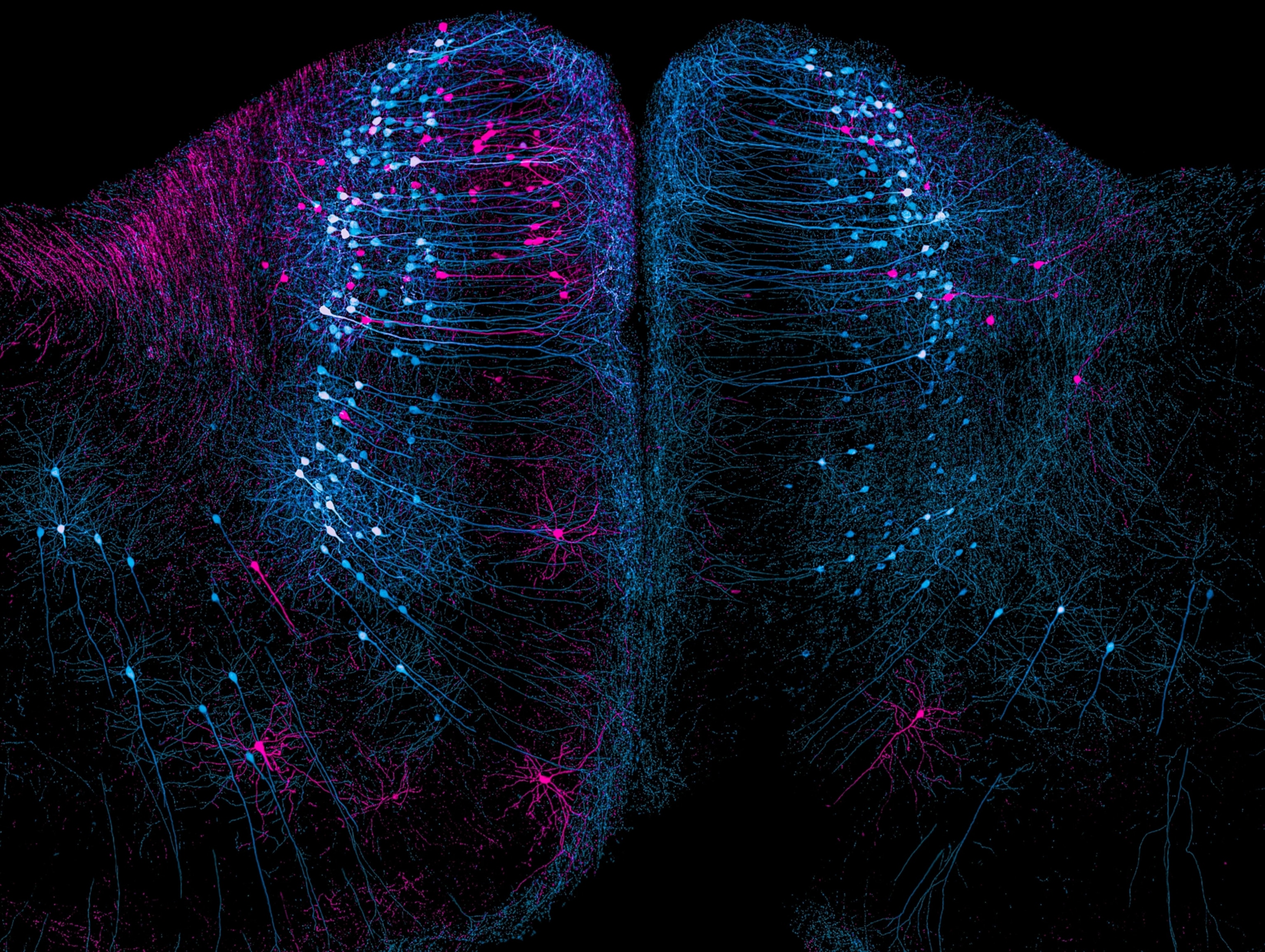
A microscopic view of bundles of mouse cortical nerve cells, labeled with fluorescent pink or blue proteins depending on which visual field activates which neurons (blue corresponds to the upper field of vision, while pink corresponds to the lower field of vision)
Our brains are hardwired to react predictably to certain stimuli. When we see objects above us or sense them behind us, we react more apprehensively than when we observe something at eye level. The brain cells seen firing in a rodent’s brain here are helping scientists understand how the brain processes visual information.
Photograph by Nikky Corthout & Alex Calzoni, Nikon Small World
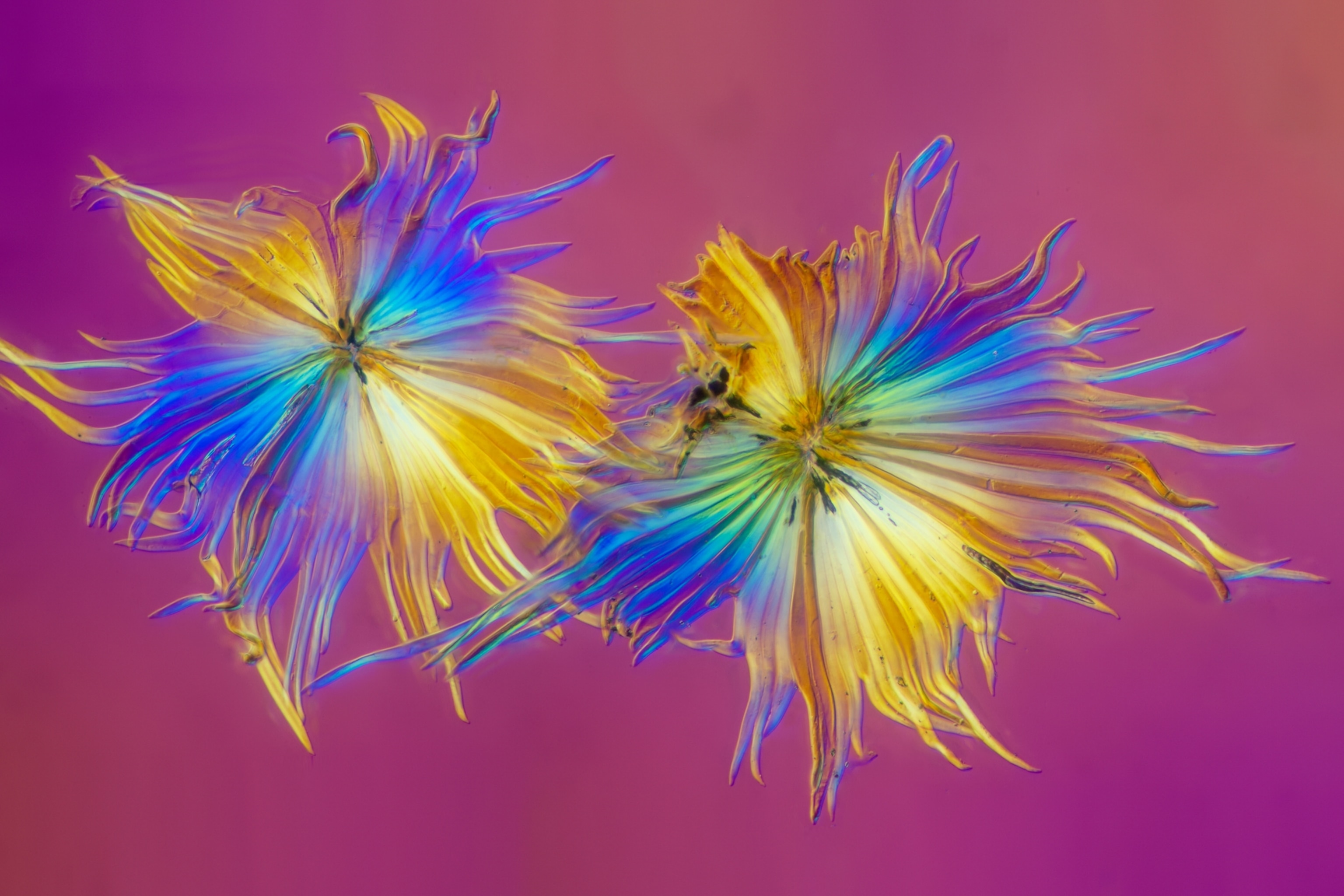
Two buckthorn trichome hairs, which look almost like two flowers with petals of varying hues of gold and blue
Leaves growing on sea buckthorn plants grow tiny hair-like fuzz called trichomes. Removed from a leaf with a razorblade, these thin, nearly transparent hairs refract light when illuminated under a microscope.
Photograph by Walter Machielsen, Nikon Small World