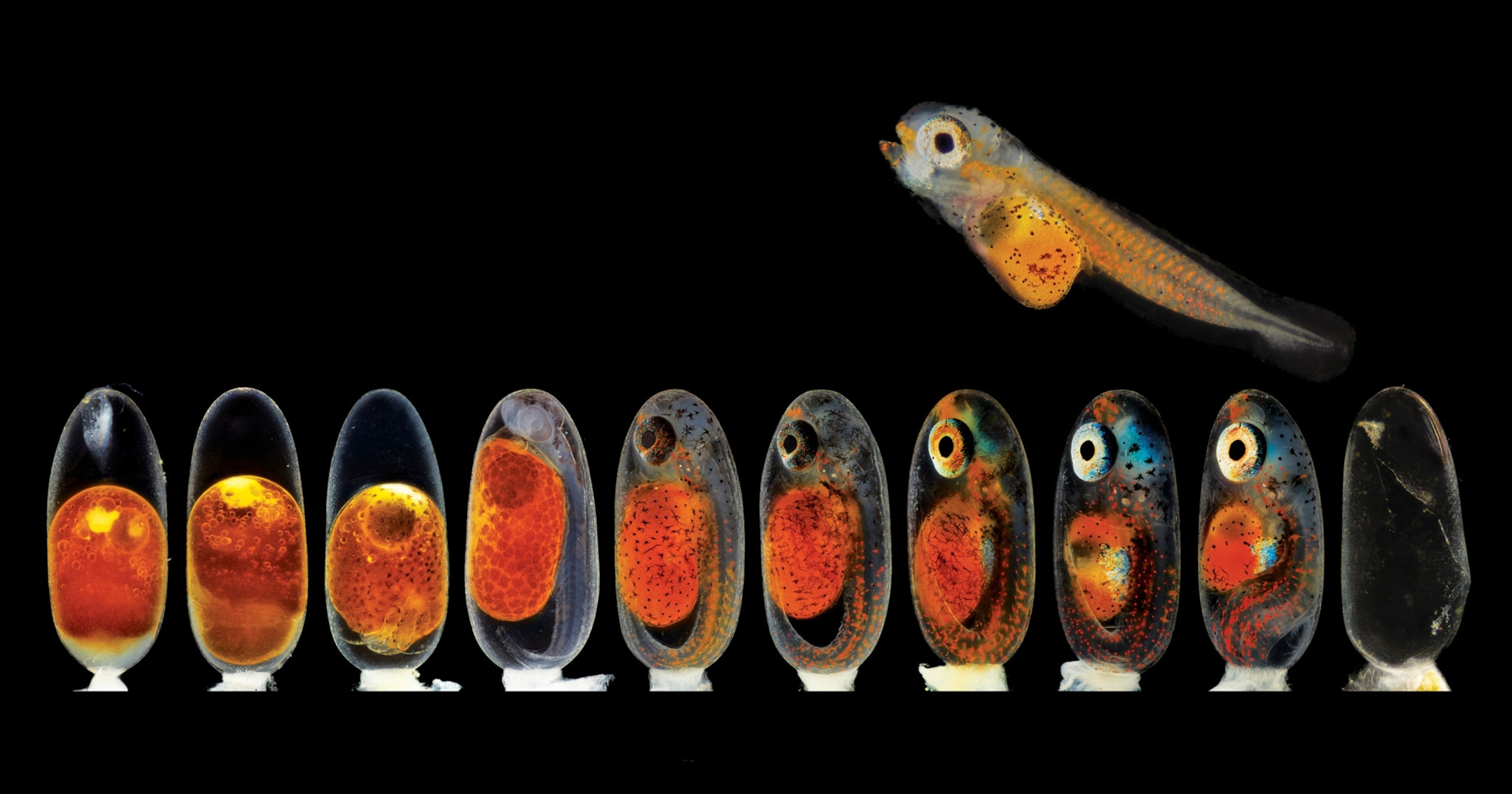
Microscopic image of clownfish embryos in several developmental stages

Revel in the tiny marvels seen through a microscope

These images, honored in Nikon’s annual Small World photo competition, are reminders of the colorful wonders invisible to the naked eye.

Nikon Small World 2021Watch a clownfish transform from a bright orange ball of cells to a small swimmer in this image from Daniel Knop of Nature and Animal Publishing House in Germany. The fishy development is fast: The entire process commonly only takes about a week from fertilization to freedom.
Daniel Knop
ByMaya Wei-Haas
September 13, 2021
8 min read

Color, texture, and shape sublimely intermingle in the world outside my window. Mottled green vines grow in a tangle punctuated by shiny red orbs of cherry tomatoes. Scalloped leaves drift to the ground as the wind rustles the tree branches. But gazing at this tableau, I know another visual feast hides in plain sight: the world of the tiny.

From spiky grains of pollen to the golden scales of a butterfly’s wing, the smallest details are easy to overlook. So every year Nikon’s Small World Photomicrography Competition reminds us of that diminutive beauty. Now in its 47th year, the competition received 1,900 entries from 88 countries from people seeking to visualize the details often invisible to the naked eye.

Nikon’s competition honors the rapidly advancing technology of photomicrography. The tools and techniques available have advanced by leaps and bounds since the father-and-son duo Hans and Zacharias Janssen, both Dutch spectacle-makers, created the first microscope in 1590. That discovery opened up a new world to explore—right down to the plaque on humans teeth, which Antony van Leeuwenhoek described in 1683 as white matter “thick as if ’twere batter.”

This year’s contest entries reveal extraordinary details of some surprisingly ordinary things, from the vibrant glow of backlit sand to the plume-like antennae atop a midge’s head. All this year’s winners are available for viewing on Nikon’s website. And here, National Geographic photo editor Todd James selected the images that captured his imagination among this year’s full crop of honorees.

“For me, this contest is less about who won than a wonderful reminder of how miraculous nature is at every scale,” James says.
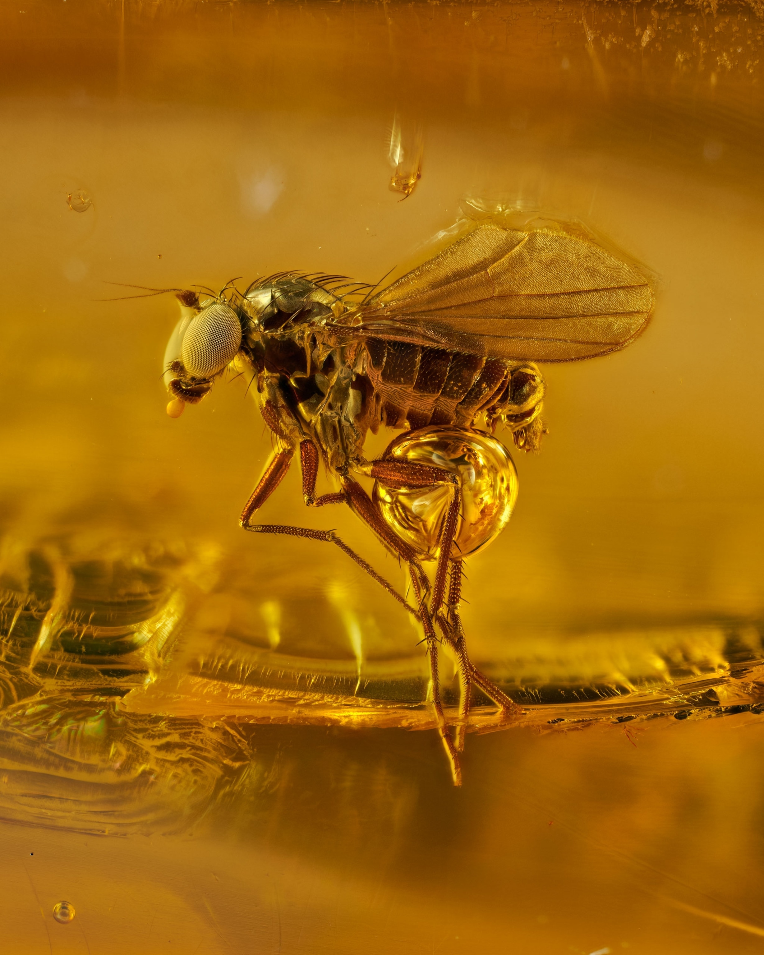
Microscopic image of a 40 million year old gnat in amber
While this gnat appears to be caught mid-flight, the hapless critter is actually frozen in time, enveloped by tree resin some 40 million years ago. The tiny treasure, shown in this image by photographer Levon Biss, hails from the Baltic region of northern Europe, which boasts the largest amber deposit yet found.
Levon Biss
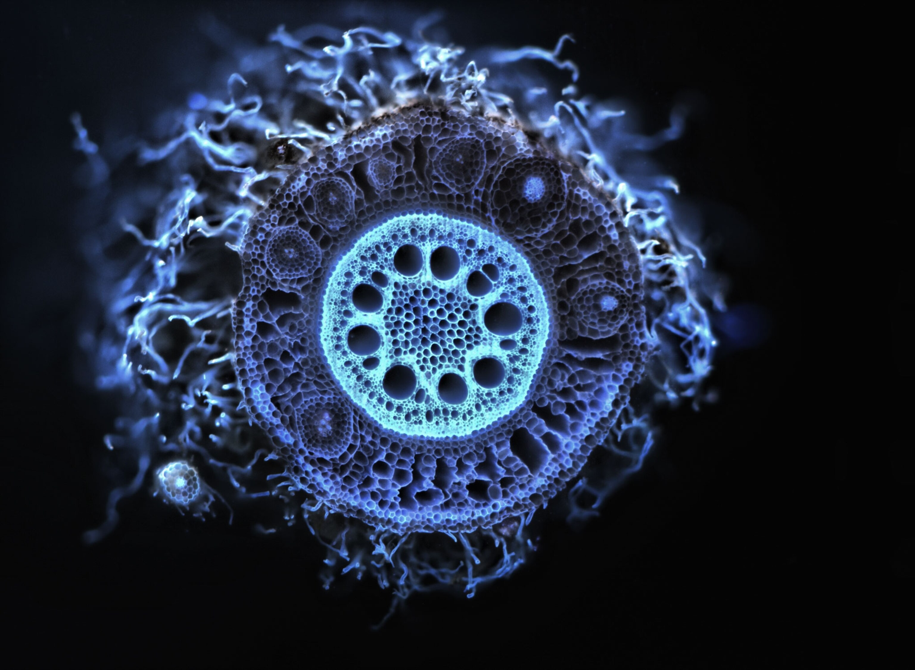
Microscopic image of a root cross section
What appears to be a wiggly sea creature is actually the sliced crown of the root of a pearl millet plant, shown here at five times the magnification. University of Nottingham’s Dylan Jones made the image with a revolutionary new method known as laser ablation technology, in which laser pulses are used to vaporize the root surface, exposing a clean view of the plant’s internal structures for photography.
Dylan Jones
Microscopic image of a butterfly wing
At 20-times magnification, the vein and scales of a Morpho didius butterfly wing begin to look like sculptures wrought in gold and bronze. The fine structures etched into the wings’ scales cause incoming light to diffract and interfere, which gives the butterfly’s wings a metallic blue color to the human eye. Taken by French photographer Sébastien Malo, this image was awarded 10th place in this year’s contest.
Sébastien Malo
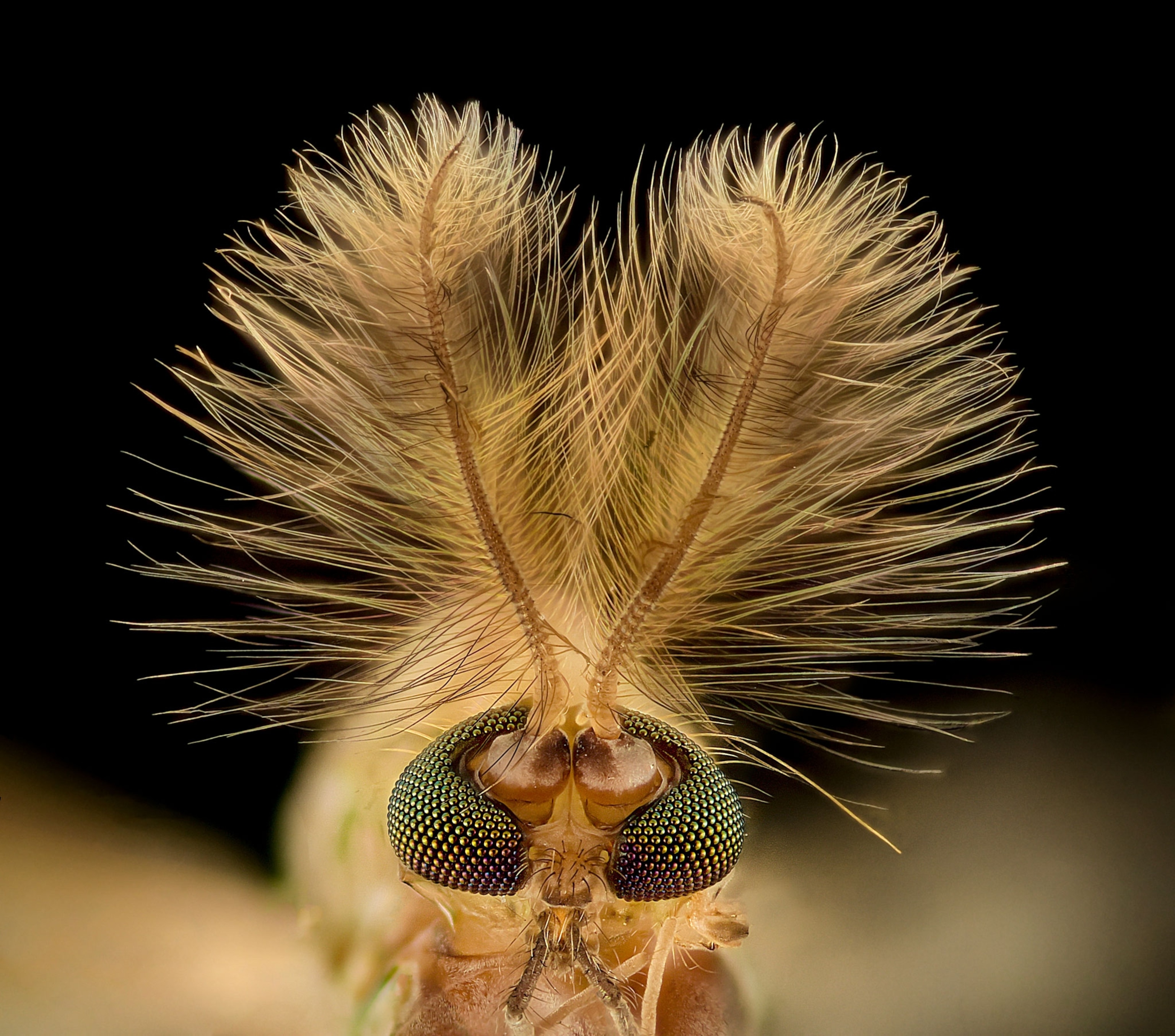
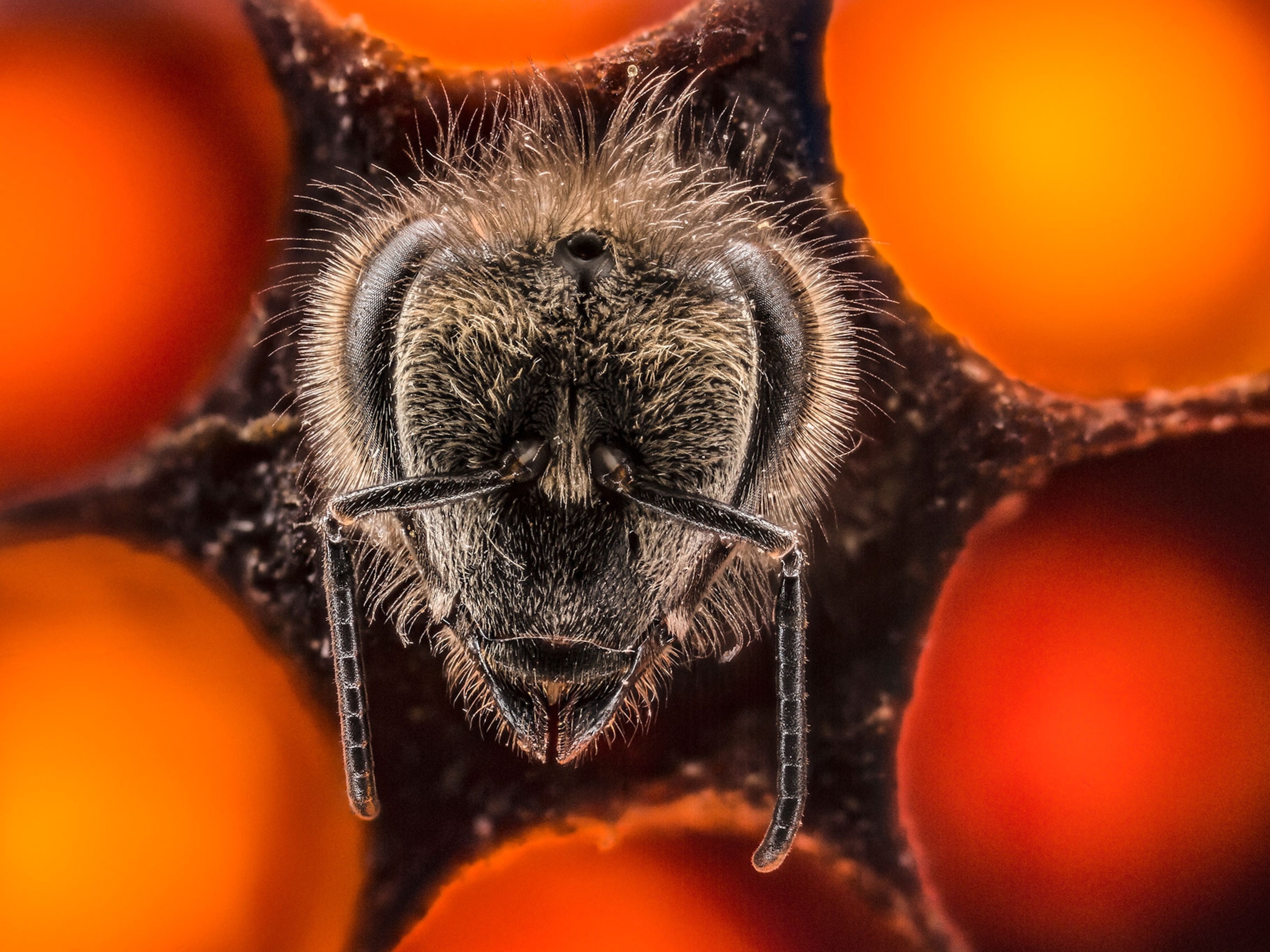
Microscopic image of a midge
A pair of plume-like antennae protrude from the head of a midge in this image made by Erick Francisco Mesén. The ornate headgear differs between male and female midges, perhaps allowing the bugs to tune into different sound frequencies. The plumes’ precise purpose is unknown, but scientists speculate that similar plumes on male mosquitoes—the blood-sucking relatives of the midge—help them spot females flying nearby.
Dr. Erick Francisco Mesén
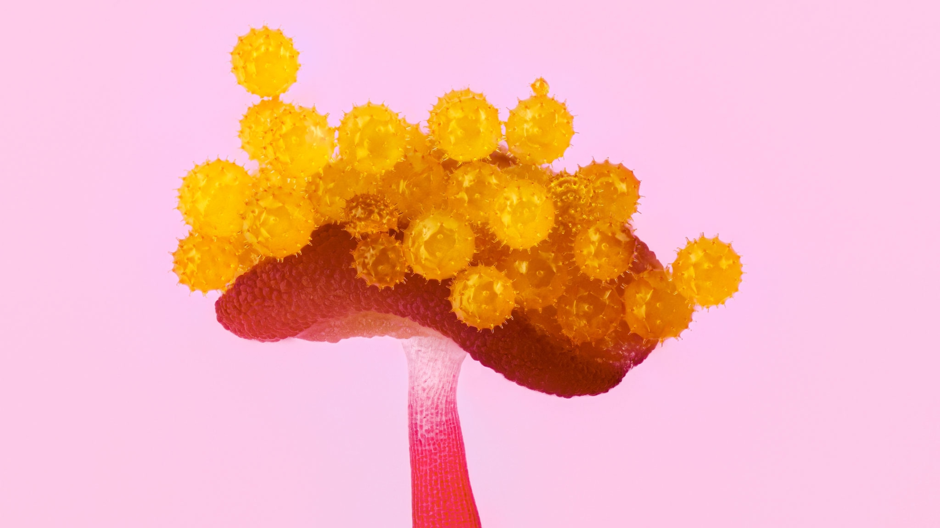
Microscopic image of a hibiscus anther
Vibrant yellow grains of pollen seem precariously balanced atop the tip of a hibiscus anther in this image made by photographer Guillermo López López. The anther, shown here at 10-times magnification, both produces and disperses pollen and is part of the plant’s male reproductive organ, the stamen.
Guillermo López López
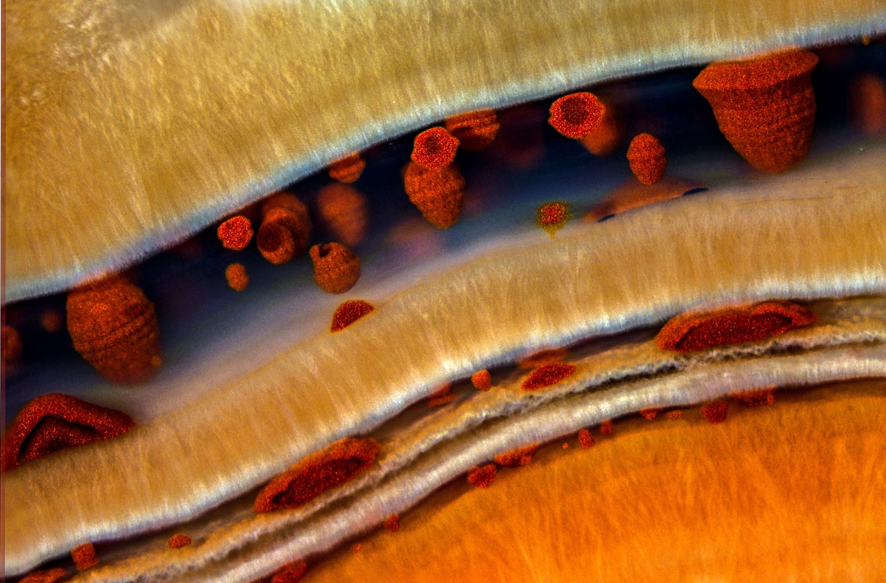
Microscopic image of Fairburn agate
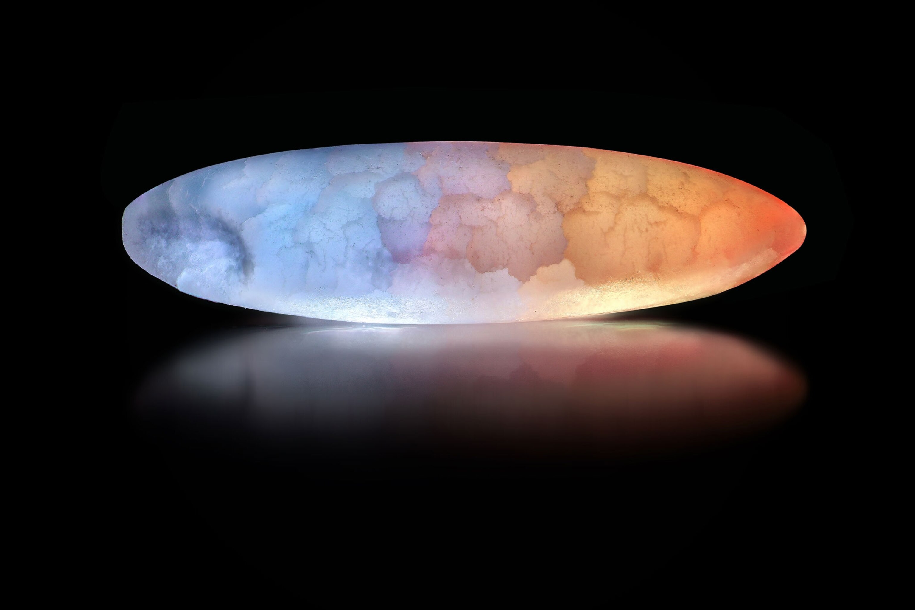
At 63-times magnification, the mineral agate looks like abstract art, as seen in this image taken by photographer Douglas Moore of the University of Wisconsin. The vibrant mineral forms from the crystallization of silica-rich waters as it percolates through rocky voids, most often in freshly cooled lava flows. Variations in the crystal size, structure, and composition define the mineral’s characteristic layers.
Douglas Moore
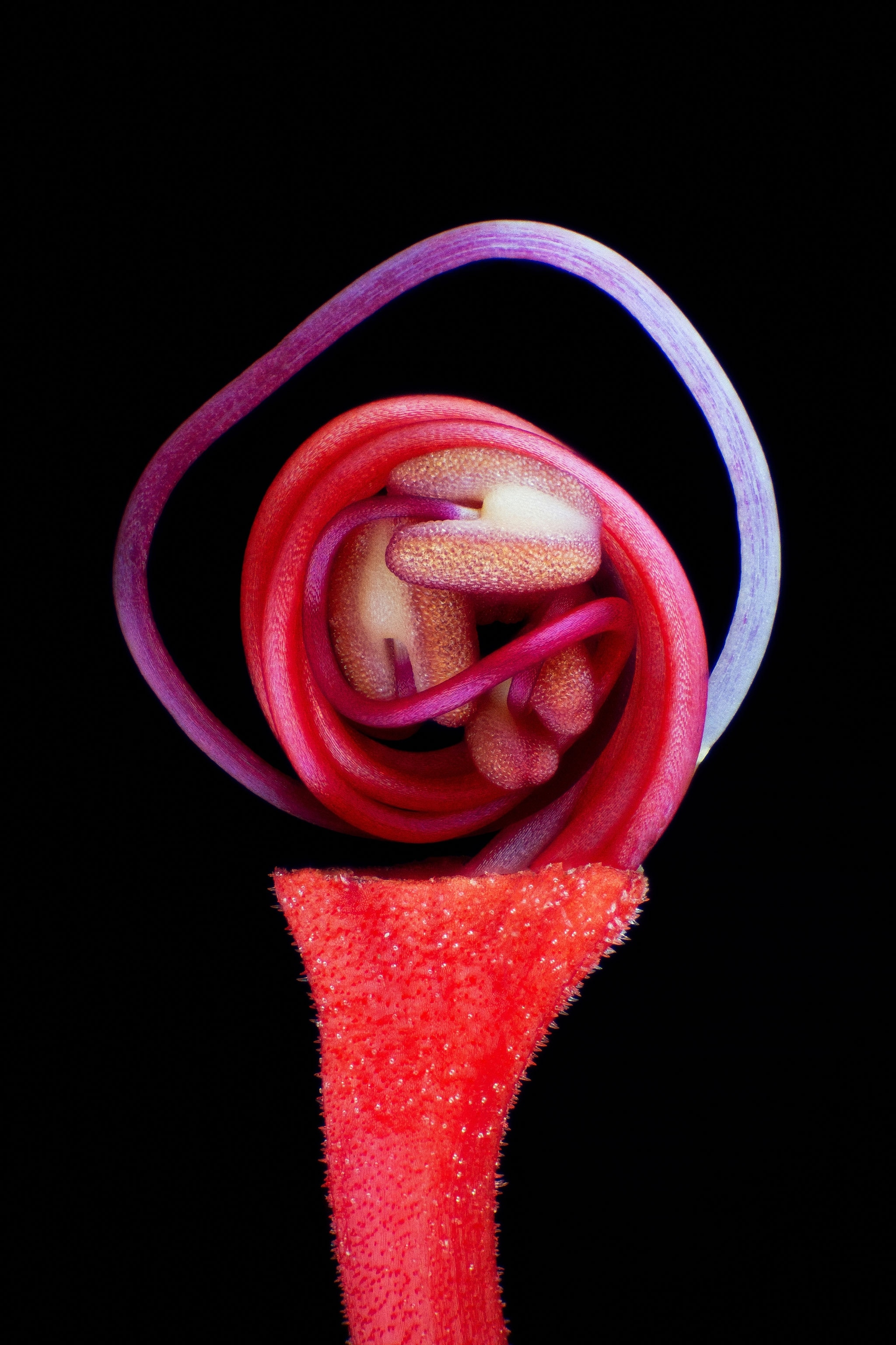
Microscopic image of a flower bud
Ever wonder what lies beneath a flowering bud’s petals? José Almodóvar, a biologist at the University of Puerto Rico, took this image of Clerodendrum paniculatum, commonly known as the “pagoda flower,” denuded of its petals, which reveals the bud’s lanky magenta pistil and stamens still curled up tight.
Jose R. Almodovar
Microscopic image of a brine shrimp
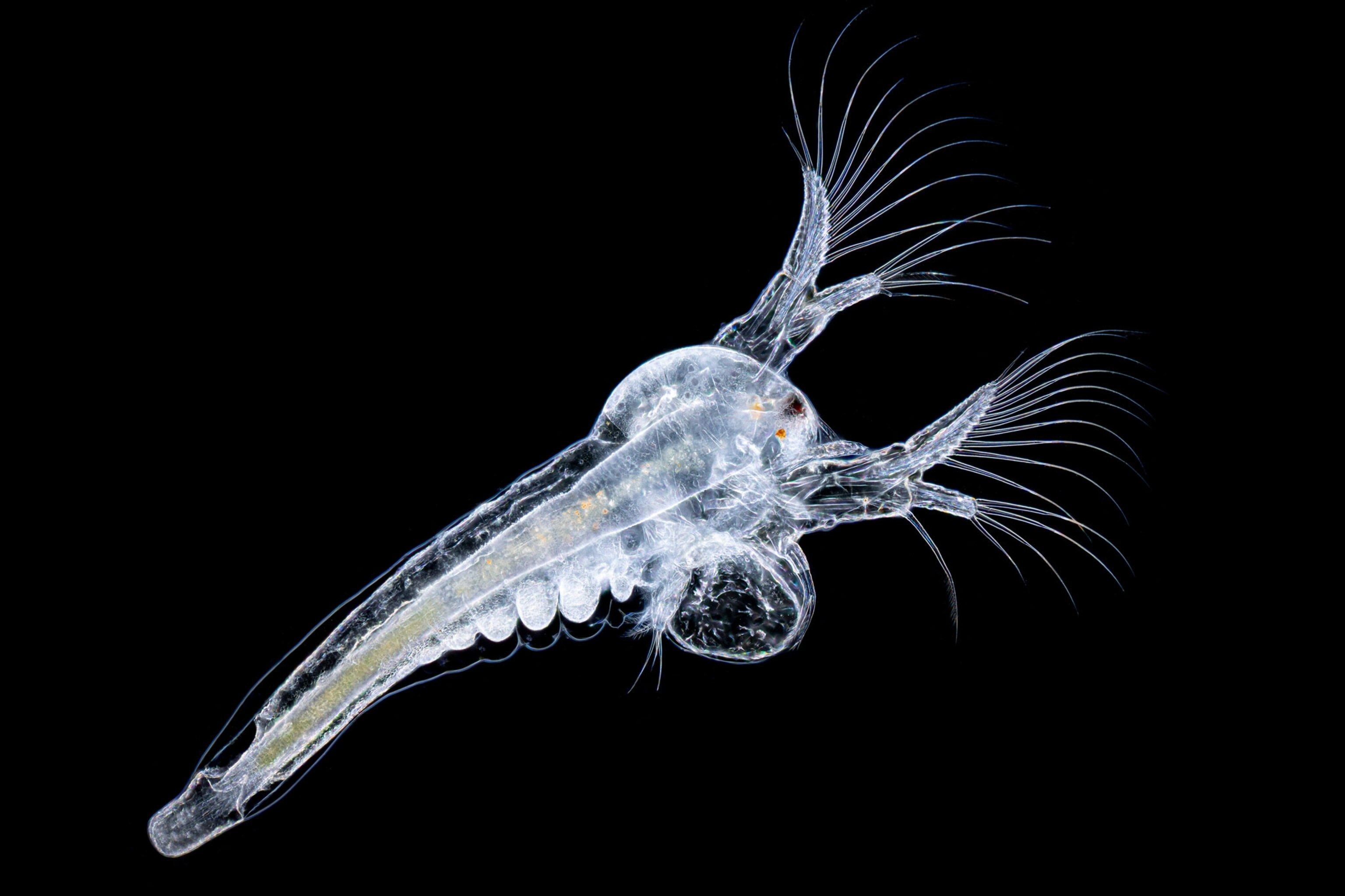
This tiny translucent brine shrimp is a mere four days old, shining in glowing contrast against a sea of black in this image by photographer Waldo Nell. Shown here at 10-times magnification, brine shrimp lay dormant embryos known as cysts that can be stored for long periods of time, making them an ideal food source in aquaculture. Many people may be familiar with one variety of brine shrimp under a more adorable name: sea monkeys.
Waldo Nell
Microscopic image of a mango seed weevil
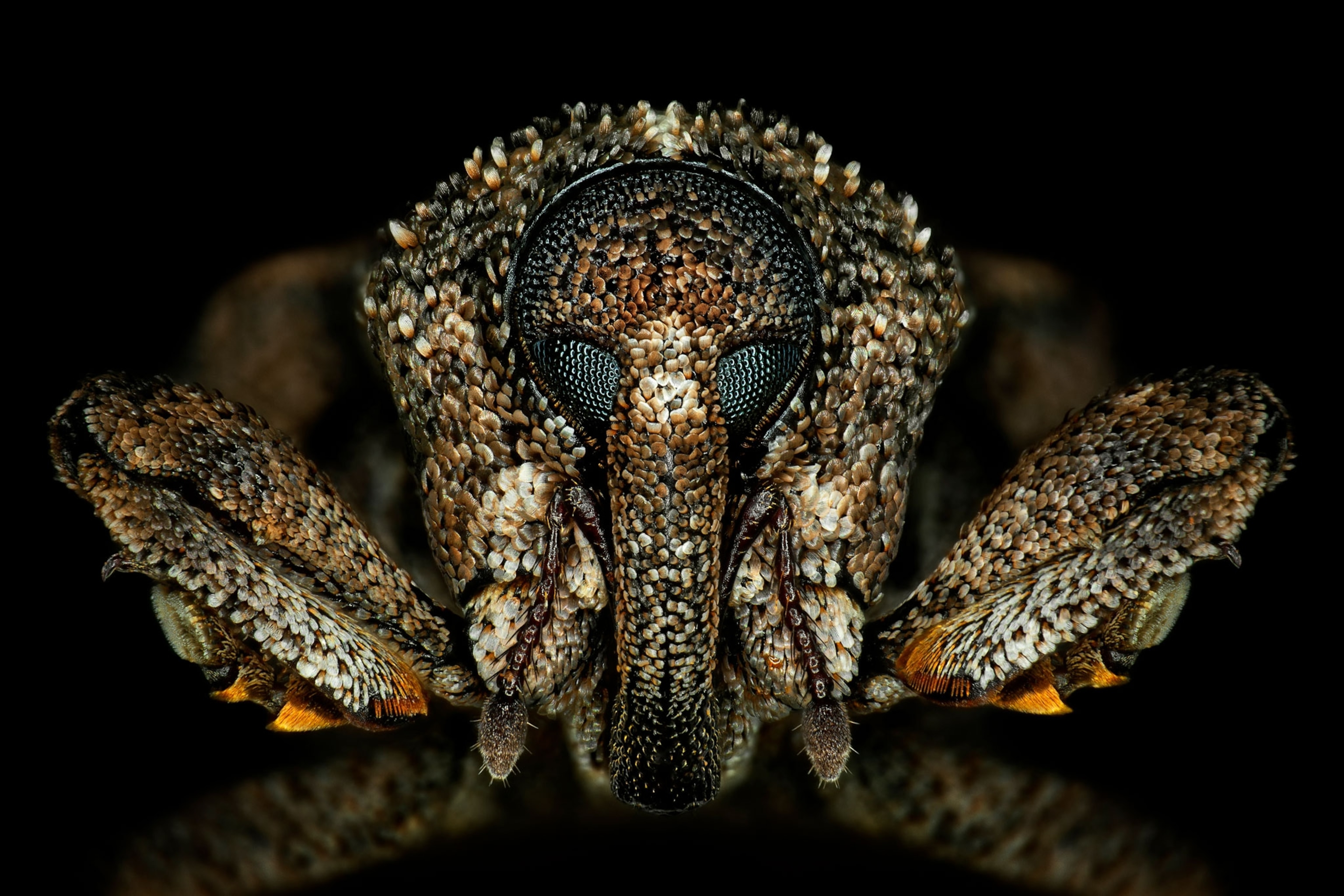
While this face may resemble the beasts that haunt your dreams, it prefers mangoes to humans. The babies of the mango seed weevil burrow into the fruit’s giant seed, where they feast until the larvae turn to adults, one of which is shown in this image by Antoine Franck of France’s Center for International Research and Cooperation.
Antoine Franck
Microscopic image of desert sand
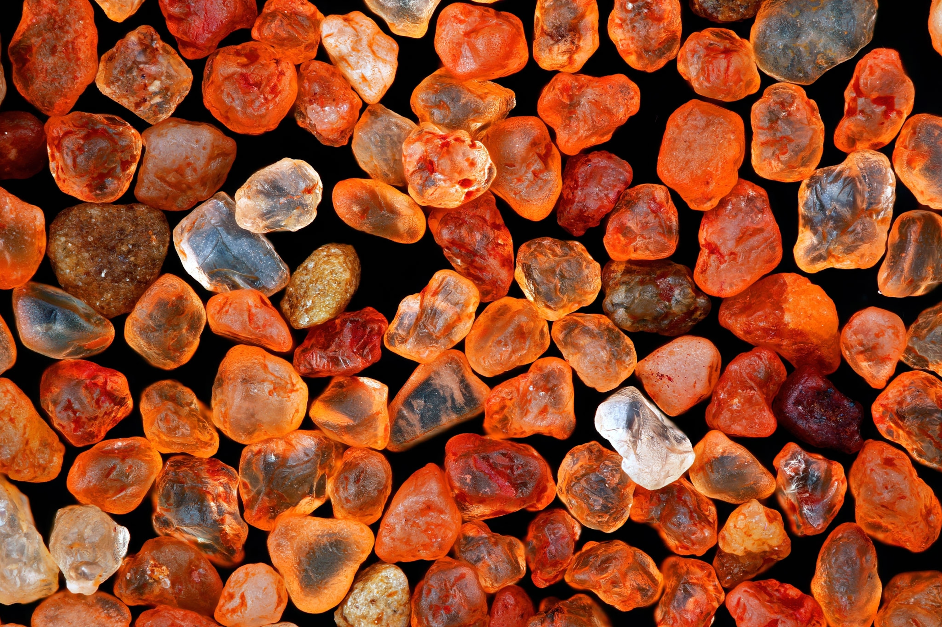
These glowing rocks may resemble the infinity stones of the fictional Marvel Universe, but they’re actually bits of sand at 10-times magnification in an image made by photographer Xinpei Zhang. Collected from the Namib Desert in southern Africa, the sand gets its brilliant color from an abundance of iron.
Xinpei Zhang
Microscopic view of a grain of rice
This grain of rice is surprisingly soothing to gaze upon as light highlights each crack and blemish in its starchy surface, shown in this image by photographer Roni Hendrawan at 20-times magnification.
Roni Hendrawan
Microscopic image of a sleeping cukoo wasp
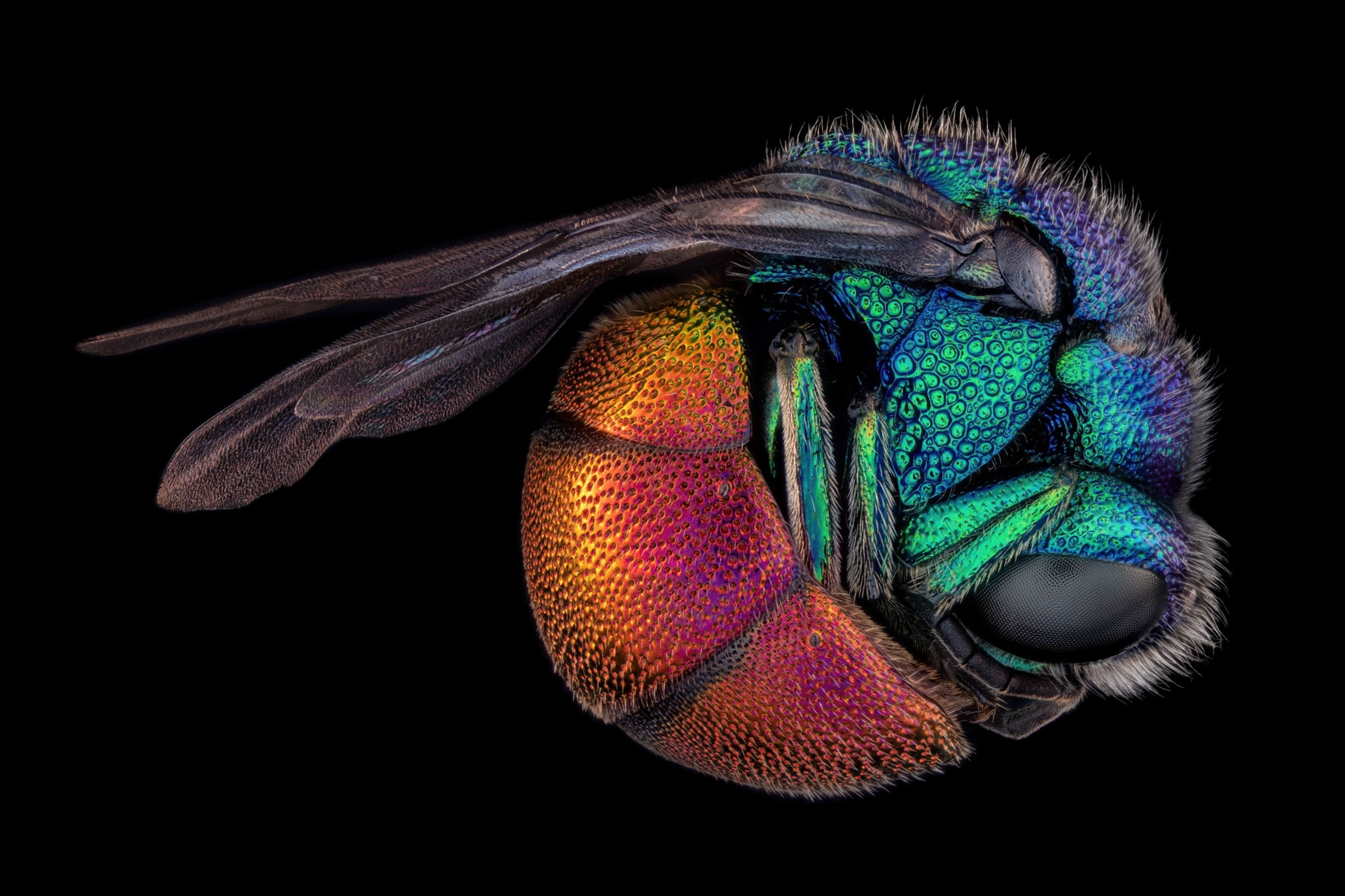
Curled in an iridescent ball, a sleeping cuckoo wasp looks serene in this image from insect photographer Thorben Danke. Don’t let the calm pose fool you: Similar to the bird that shares its name, the cuckoo wasp lays its eggs in the nests of other insects. The hatched larvae will then feed on the nest-builder’s babies or starve them to death by devouring stored food.
Thorben Danke, Nikon Small World Contest

Michael Greshko contributed reporting to this story.

LIMITED TIME OFFER

Discover More, Spend Less
With new subscriber-exclusive stories published daily and complete archive access, your opportunities to explore are endless!